Ultrasound
Make an Appointment
Online Schedule

A Better Ultrasound Experience
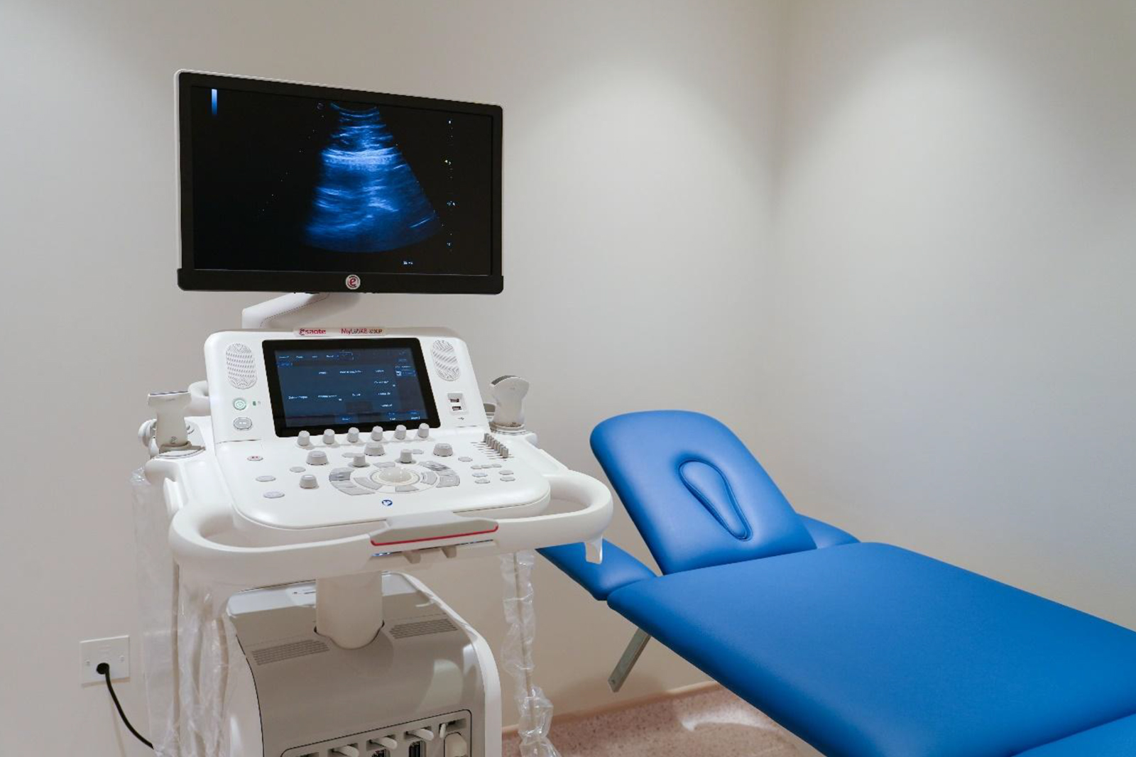
Our Center has the Ultrasound Esoate MyLab X8 from Italy.
What is an Ultrasound?
Ultrasound imaging uses high-frequency sound waves to produce pictures of the inside of the body. It does not use radiation and provides clear pictures of soft tissues that don’t show up well on X-rays. Images are captured in real time and can show the movement of the body’s internal organs. The ultrasound procedure is simple and painless. A hand-held transducer emitting silent, high frequency sound waves is placed against the body and slowly passed over the area being examined. Sophisticated equipment produces images on a video monitor for the radiologist to interpret.
Prep for your exam
Abdomen Ultrasound
OB or pelvic Ultrasound
- A full bladder is required for this exam. Drink 32oz of water 90 minutes before your exam. Please do not empty your bladder.
Ultrasound (Obstetrical/GYN, Abdominal & Vascular)
Ultrasound imaging uses high-frequency sound waves to produce images of the inside of the body. It does not use radiation and provides clear pictures of the soft tissues that don’t show up well on X-rays. Ultrasound is often used to evaluate fetus during pregnancy, and can also be used to examine the chest, abdomen and blood vessels.
Ultrasound Scanning:
- Pelvic Ultrasound Imaging
- Transabdominal Ultrasound
- Transvaginal Ultrasound
- Transrectal Ultrasound
- Obstetric Ultrasound Imaging
- Carotid & Abdominal Aorta Ultrasound Imaging
- Liver Ultrasound
- Renal Ultrasound
- Vascular Ultrasound
- Thyroid Ultrasound
What you should know Answers to frequently asked questions.
An ultrasound shows the details of structures in the abdomen. It can show features like the size and movement of organs, cysts or growths, or fluid collections. An ultrasound of the abdomen is most often done to:
Certain exams require an empty stomach or full bladder for better visibility of the organ. You may be advised to:
You will be positioned on a table. A gel will be placed over the area that will be checked. The gel helps the sound waves travel from a wand to your body.
The ultrasound machine has a hand-held wand. The wand is pushed against your skin where the gel has been applied. The wand sends sound waves into your body. The waves bounce off your internal organs and echo back to the wand. The computer can convert echoes into images on a screen. The images on the screen are examined by the radiologist. A photograph of them may be taken.


